The Microbiology Laboratory is one of the few laboratory units that operates largely independently of fully automated analyzers and advanced automated systems. Due to its reliance on hands-on processing of clinical specimens and definitive diagnostic interpretation, this department requires continuous daily supervision by a qualified specialist to ensure that all diagnostic variables are carefully controlled and that results remain accurate and reliable under established quality control programs.
The Microbiology Department includes dedicated sections for microbial culture, media preparation, incubation, and sterilization. Sterile culturing of body fluids and secretions on appropriate microbiological media, as well as direct bacteriological examinations and staining procedures, are routinely performed to identify causative pathogens. After pathogen identification, an antibiogram (antimicrobial susceptibility test) is conducted to determine the sensitivity of the isolated organism to various antibiotics.
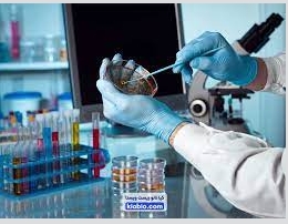
Various laboratory methods are used in microbiological testing to differentiate and identify microbial agents. A comprehensive laboratory guide is available for detailed procedures and protocols related to these tests.
Importance of Culture Media in Microbiological Testing
In many cases, bacterial species cannot be identified solely based on morphological characteristics. Therefore, microorganisms must be cultured on artificial media to isolate them and evaluate their bacteriological properties. When more than one microorganism is present in a culture, they must be separated to obtain a pure culture for accurate study.
Three fundamental principles must be observed in microbial investigation:
-
Preparation of appropriate culture media
-
Elimination of contaminating microorganisms through sterilization
(Since microorganisms are ubiquitous and may contaminate materials used in media preparation, culture media must be completely sterile before inoculation.) -
Proper cultivation and isolation of microorganisms from mixed samples
Gram stain in Microbiological Testing
Gram staining is one of the most widely used staining techniques in microbiology. It was first developed by the Danish bacteriologist Hans Christian Gram. Based on this method, bacteria are classified into two groups: Gram-positive and Gram-negative.
The staining outcome depends on the bacterium’s ability to retain the primary dye, which is determined by the structure of its cell wall. Gram-positive bacteria possess a thicker peptidoglycan layer in their cell wall, allowing them to retain the crystal violet stain, whereas Gram-negative bacteria have a thinner peptidoglycan layer and do not retain the primary stain after the decolorization step.
Microbiological Laboratory Guide
The laboratory guide (89 pages, in Persian) includes detailed descriptions of various culture media, preparation methods, diagnostic procedures, Gram staining techniques, and additional microbiological tests.
Some of the topics covered include:
-
Bile Esculin Agar
-
CAMP test
-
Citrate test
-
DNase Test Agar
-
Kligler Iron Agar
-
Methyl Red test
-
Phenylalanine deaminase test
-
PYR test
-
Bacitracin and SXT sensitivity testing
-
TCBS agar
-
Triple Sugar Iron test
-
Voges–Proskauer test
-
Decarboxylation tests
-
Salt tolerance testing
-
Preparation of various culture media, reagents, and stains
-
Technical procedures for preparation and quality control of culture media
-
Loop volume determination methods